Secondary Structure
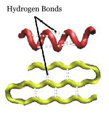
The secondary structure of a protein refers to stable local folding of portions of the protein involving hydrogen bonding between backbone atoms. The two most common secondary structures are the alpha helix and the beta pleated sheet.
The secondary structure is maintained by hydrogen bonds between the backbone atoms. These form between the H of the N (amide hydrogen) and the O of C=O (carbonyl oxygen).
Alpha Helices
Alpha helices form a right-handed corkscrew within a protein. Alpha helices can range in length from very short (involving only a few amino acids) to very long (up to over seventy amino acids). Click on the buttons below to see alpha helices (colored red) within a protein.
Alpha helices are pictured in the proteins insulin and beta globin and can be identified by the color red. Note they look like a corkscrew. (2HIU.pdb, 1A3N.pdb)
Insulin 2HIU.pdb
Beta Globin 1A3N.pdb
Digging Deeper
Count the number of hydrogen bonds and the number of amino acids in the highlighted alpha helix.
Alpha Helix Note the hydrogen bonds in an alpha helix of beta globin. (1A3N.pdb)
 Determine the ratio of hydrogen bonds per amino acid.
Where are the amino acid sidechains located in the alpha helix?
Beta Pleated Sheets
The second common secondary structure is the beta pleated sheet, which consists of two or more beta strands. The backbone of a beta strand bends back and forth like a pleat (hence the name). Alternating sidechains are on opposite surfaces of the beta sheet. Hydrogen bonds connect adjacent strands.
Adjacent beta strands can lie in two different orientations. If the N-termini of both strands are adjacent to each other, they are said to be parallel beta strands, The hydrogen bonds between parallel beta strands zig zag between the strands, much like the lace in a shoe.
If the N-terminus of one strand lies adjacent to the C-terminus of the other strand, the two strands are said to be antiparallel beta strands. The hydrogen bonds between antiparallel beta strands run parallel to one another and look like the rungs of a ladder.
A protein may contain both parallel and antiparallel beta strands, often within the same beta sheet!
Green Fluorescent Protein Beta pleated sheets look like parallel lines in the green fluorescent protein (GFP) and are colored yellow. (1EMB.pdb) GFP is the protein that makes jellyfish glow, and it has been engineered to glow various colors (blue, red, gold). Scientists sometimes insert this gene in front of a gene they are studying to understand better when and where the protein is expressed in an individual. Perhaps you have seen images of glowing mice!
Digging Deeper
Count the number of hydrogen bonds and the number of amino acids in the selected beta sheet.
Beta Sheet Look at the selected beta pleated sheet from green fluorescent protein. (1EMB.pdb)
Most of the beta strands in GFP run antiparallel to adjacent strands. Can you locate the two adjacent strands that are parallel? (Hint: Look at the hydrogen bonds.)
Determine the ratio of hydrogen bonds per amino acid.
Where are the amino acid sidechains located in the beta sheet?